
|
Overview |
||
|
Product Name |
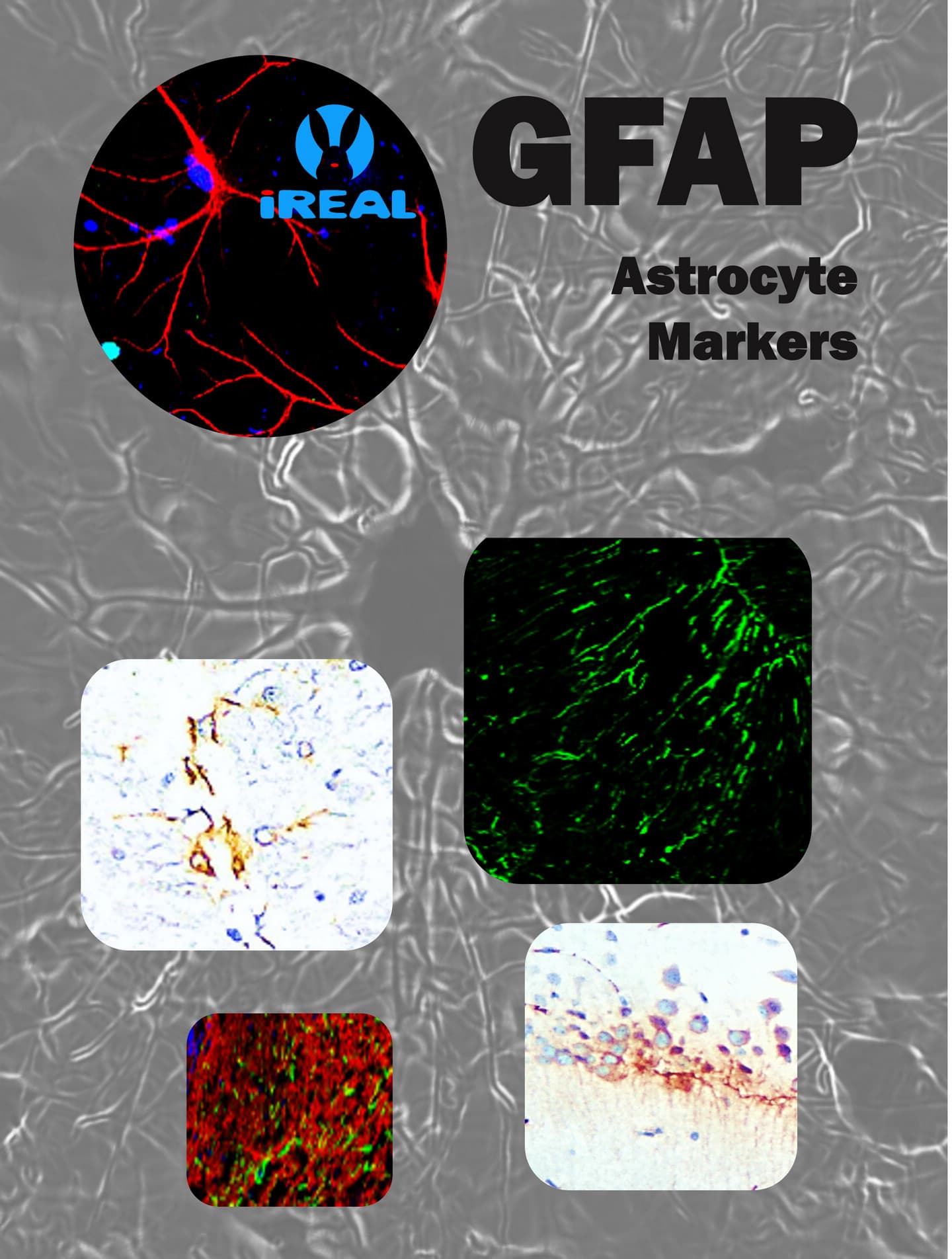
GFAP antibody |
|
|
Product Number |
||
|
Gene Description |
glial fibrillary acidic protein |
|
|
Clonality |
Polyclonal |
|
|
Host |
Rabbit |
|
|
Species Reactivity |
Human, mouse, rat |
|
|
Recommended Applications Dilutions |
Western Blot 1:1000-1:2000 Immunofluorescence 1:200 – 1:500 Immunohistochemistry (Frozen) 1:100 – 1:300 Immunohistochemistry (Paraffin) 1:100 – 1:300 |
|
|
Storage Buffer |
100mM Tris Glycine, 1% BSA, 20% Glycerol (pH7). 0.025% ProClin 300 was added as a preservative |
|
|
Concentration |
0.31 mg/ml |
|
|
Purity |
Affinity column purified |
|
|
Storage |
Store at +4°C for short term storage. Long time storage is recommended at -20°C |
|
|
Notes |
Gently mix before use. Optimal concentrations and conditions for each application should be determined by the user. |
|
|
IR15-44 anti-GFAP antibody WB image |
IR15-44 anti-GFAP antibody IHC image |
|
|
|
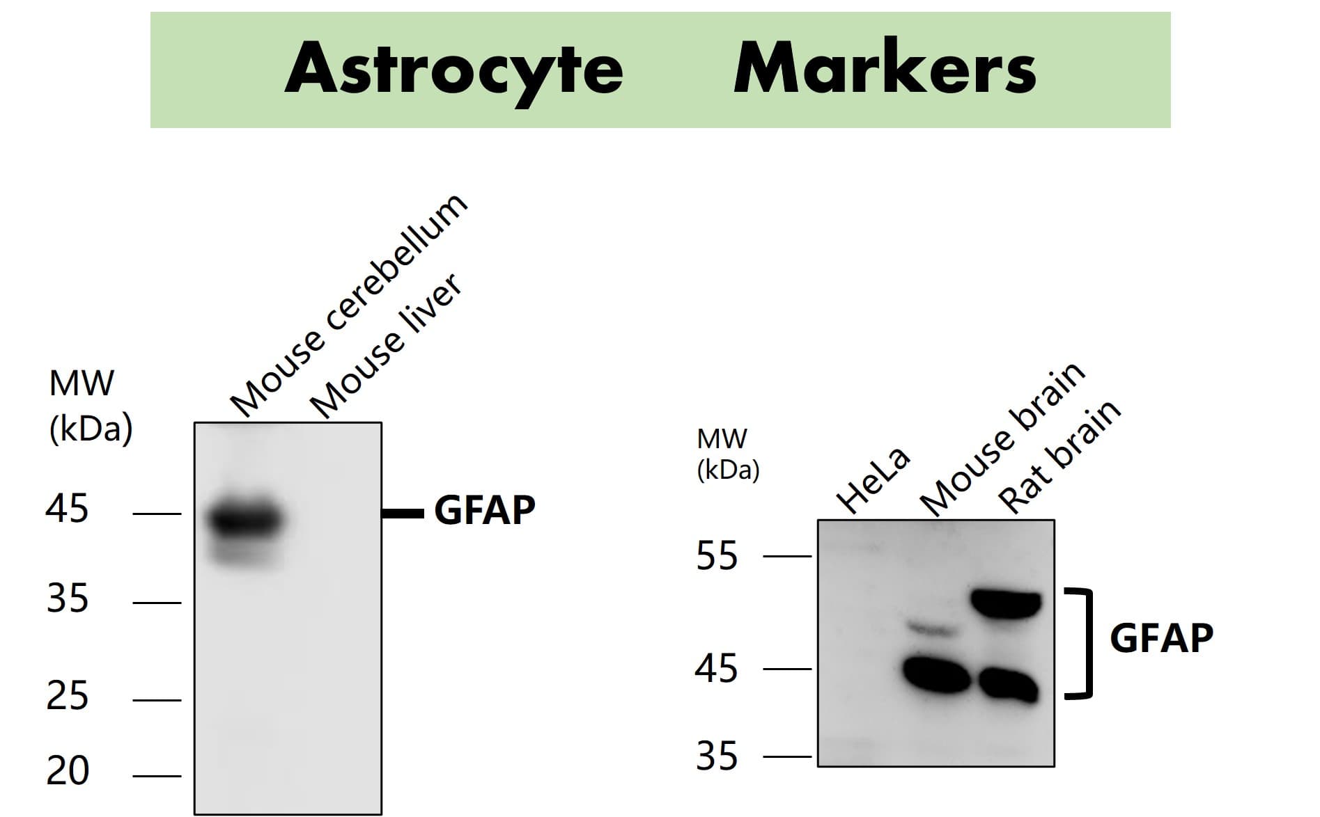
All lanes : Anti-GFAP antibody at 1/1000 dilution Lysates/proteins at 60 μg per lane This blot was produced using a 12% SDS-PAGE. Nitrocellulose was then blocked for an hour before being incubated with IR15-44 overnight at 4°C. |
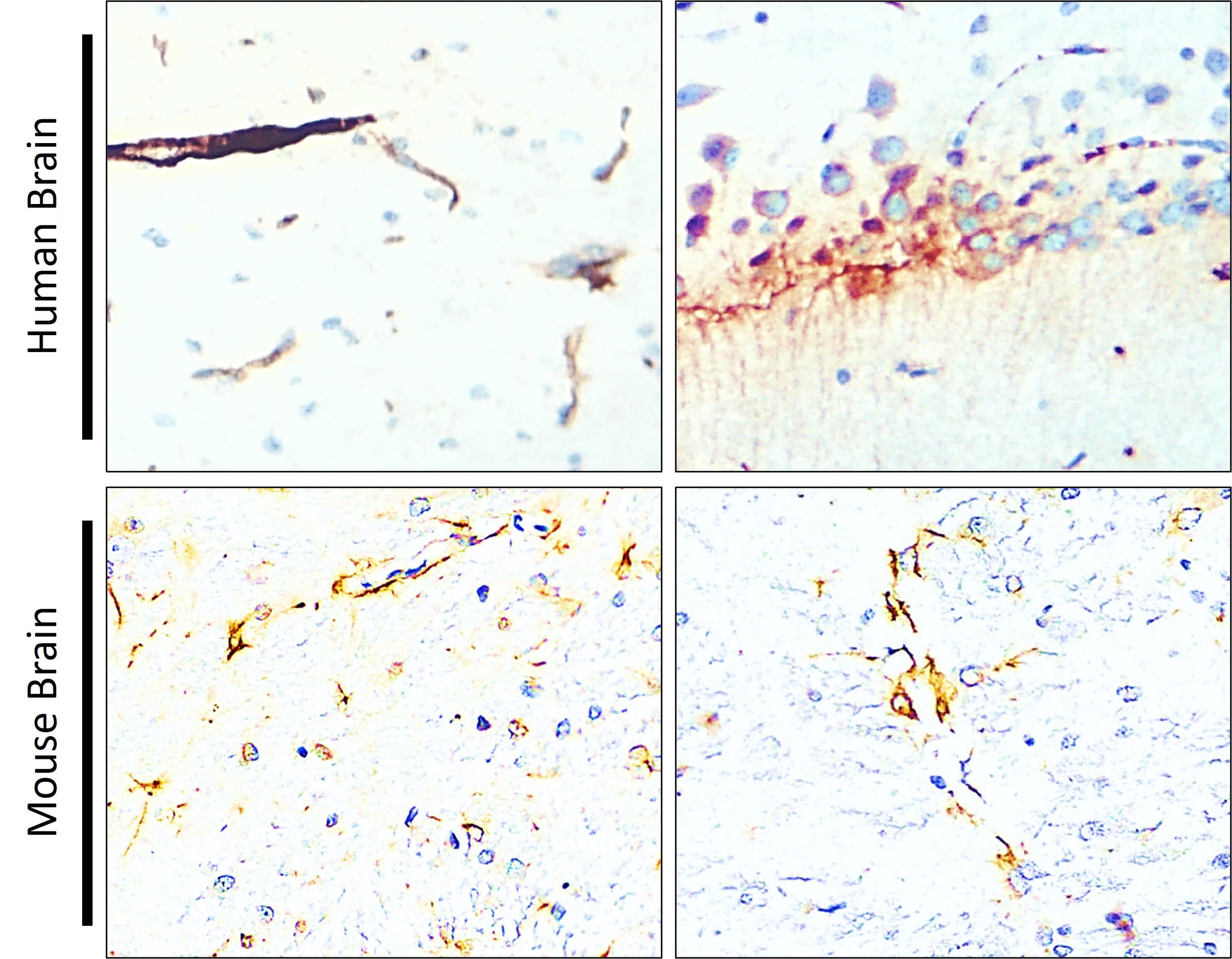
Immunohistochemical analysis of paraffin embedded Human brain tissue labeling GFAP antibody with IR15-44 at 1/100. |
|
|
|
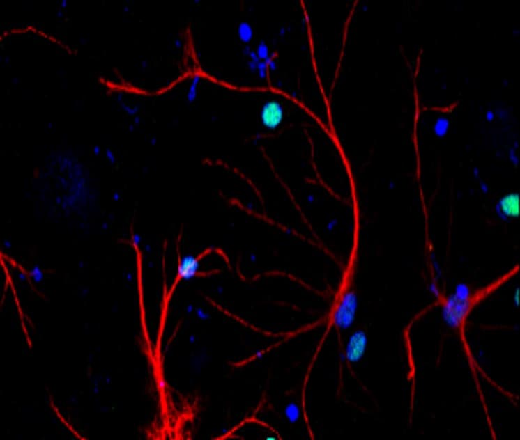
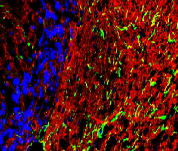
Immunofluorescent analysis. Sample : primary cortical neurons Red : GFAP (IR15-44) : 1-200 Blue : DAPI was used as the nuclear counter stain. Fixed : 4% paraformaldehyde at RT for 20 min. |
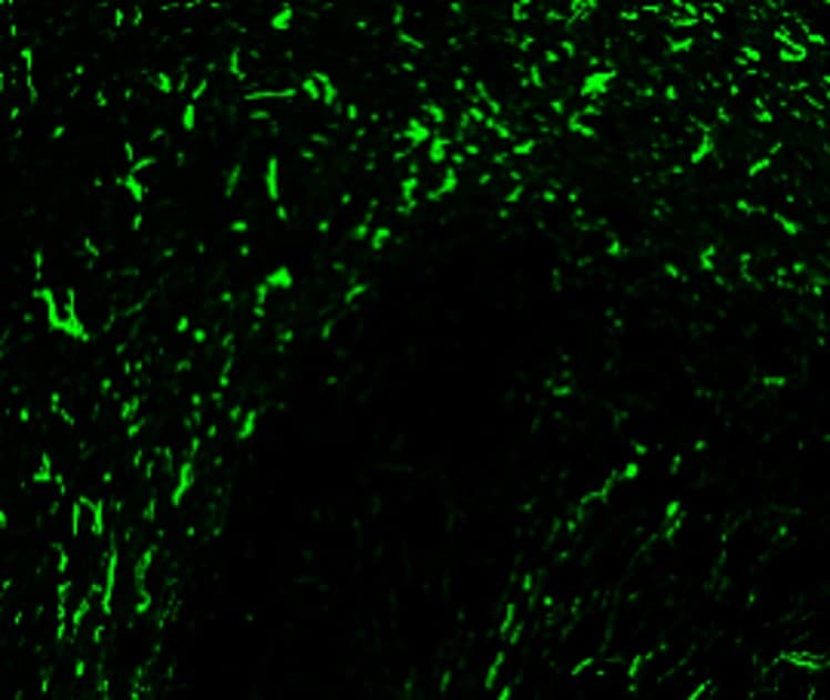
Immunohistochemical of frozen sections. Sample : mouse cerebellum. Green : GFAP (IR15-44) : 1-200 Anti-rabbit 488 : 1-500 |
|
IR15-44 anti-GFAP antibody ICC/IF image |
Customer feedback Image IR15-44 anti-GFAP antibody IHC image |
|
|
|
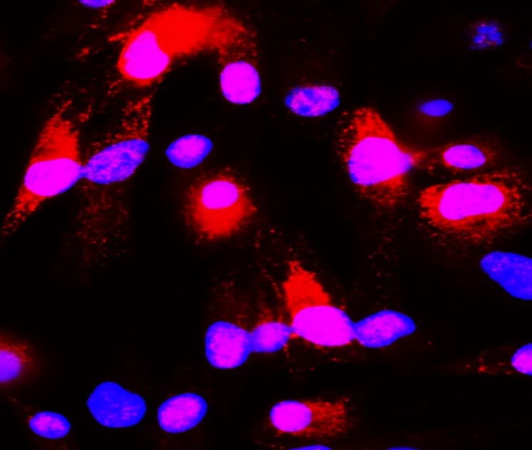
Immunofluorescence: cells were fixed with 4% paraformaldehyde for 10 min at RT, permeabilized with 0.1% NP-40 for 10 min at RT then blocked with 5% BSA for 30 min at room temperature. Cells were stained with IR15-44 anti-GFAP antibody (red) at 1:200 and 4°C. DAPI (blue) was used as the nuclear counter stain. |
Immunohistochemical of frozen sections. Sample : mouse cerebellum. Green : GFAP (IR15-44) : 1-200 Anti-rabbit 488 : 1-500 |