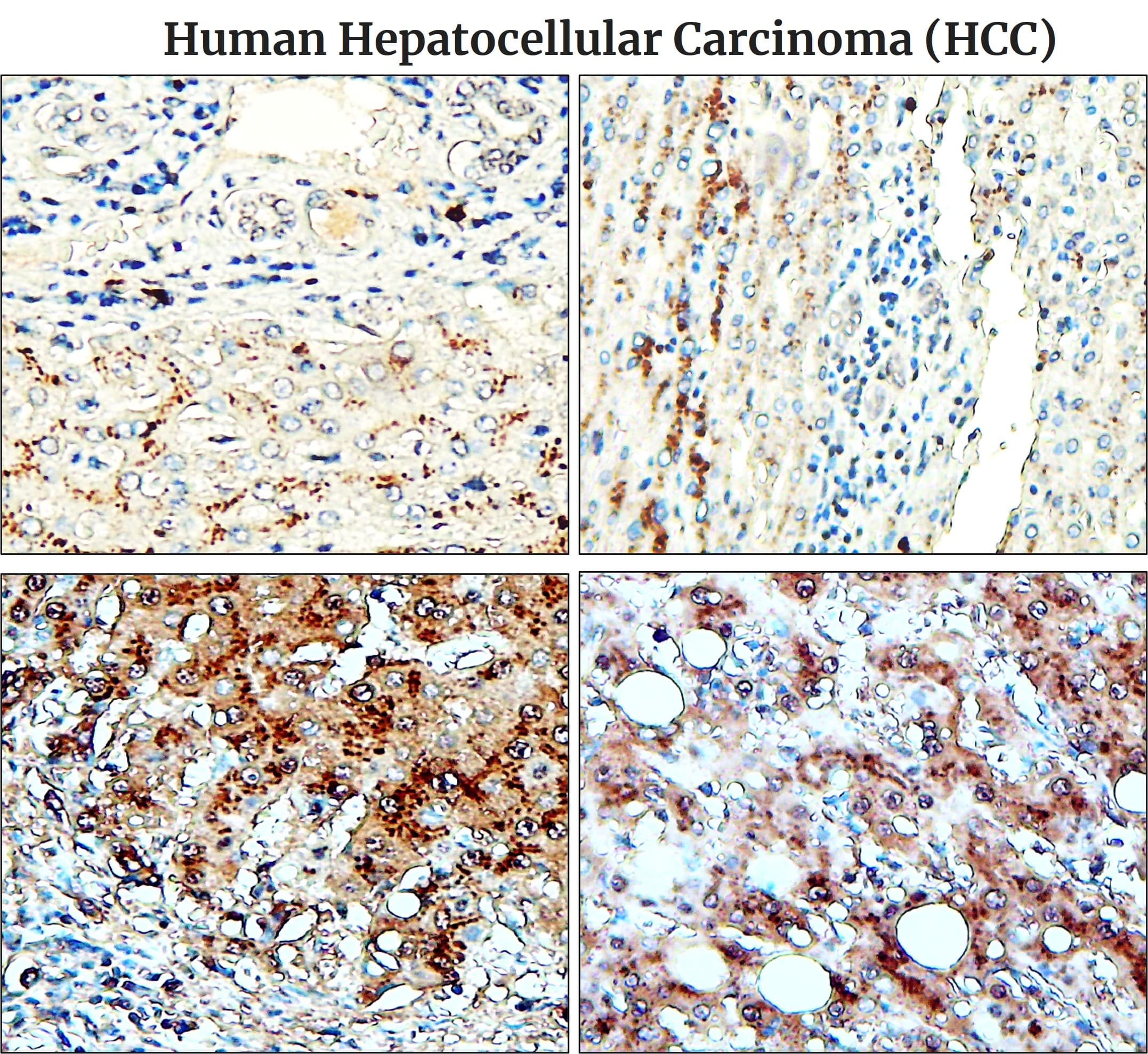
Fibronectin : FFPE Tissue patterns in human hepatocellular carcinoma ( HCC ) IR48-138 Fibronectin antibody
Fibronectin, a high molecular weight glycoprotein component of the extracellular matrix, was localized extracellularly and intracellularly in the nuclei and the cytoplasm of hepatocellular carcinomas (HCC). Nuclear fibronectin may be an expression of the DNA binding ability of fibronectin which becomes demonstrable in cells with increased membrane permeability.
|
Overview |
||
|
Product Name |
Fibronectin (C-term) antibody |
|
|
Product Number |
||
|
Gene Description |
fibronectin 1 |
|
|
Clonality |
Polyclonal |
|
|
Host |
Rabbit |
|
|
Species Reactivity |
Human, mouse, rat |
|
|
Recommended Applications Dilutions |
Western Blot 1:500 Immunofluorescence 1:200 – 1:500 Immunohistochemistry (Paraffin) 1:100 – 1:200 |
|
|
Storage Buffer |
100mM Tris Glycine, 20% Glycerol (pH7). 0.025% ProClin 300. |
|
|
Concentration |
1.5 mg/ml |
|
|
Purity |
Affinity column purified |
|
|
Storage |
Store at +4°C for short term storage. Long time storage is recommended at -20°C |
|
|
Notes |
Gently mix before use. Optimal concentrations and conditions for each application should be determined by the user. |
|
|
IR48-138 anti-Fibronectin antibody WB image |
IR48-138 anti-Fibronectin antibody IHC image |
|
|
|
|
|
|
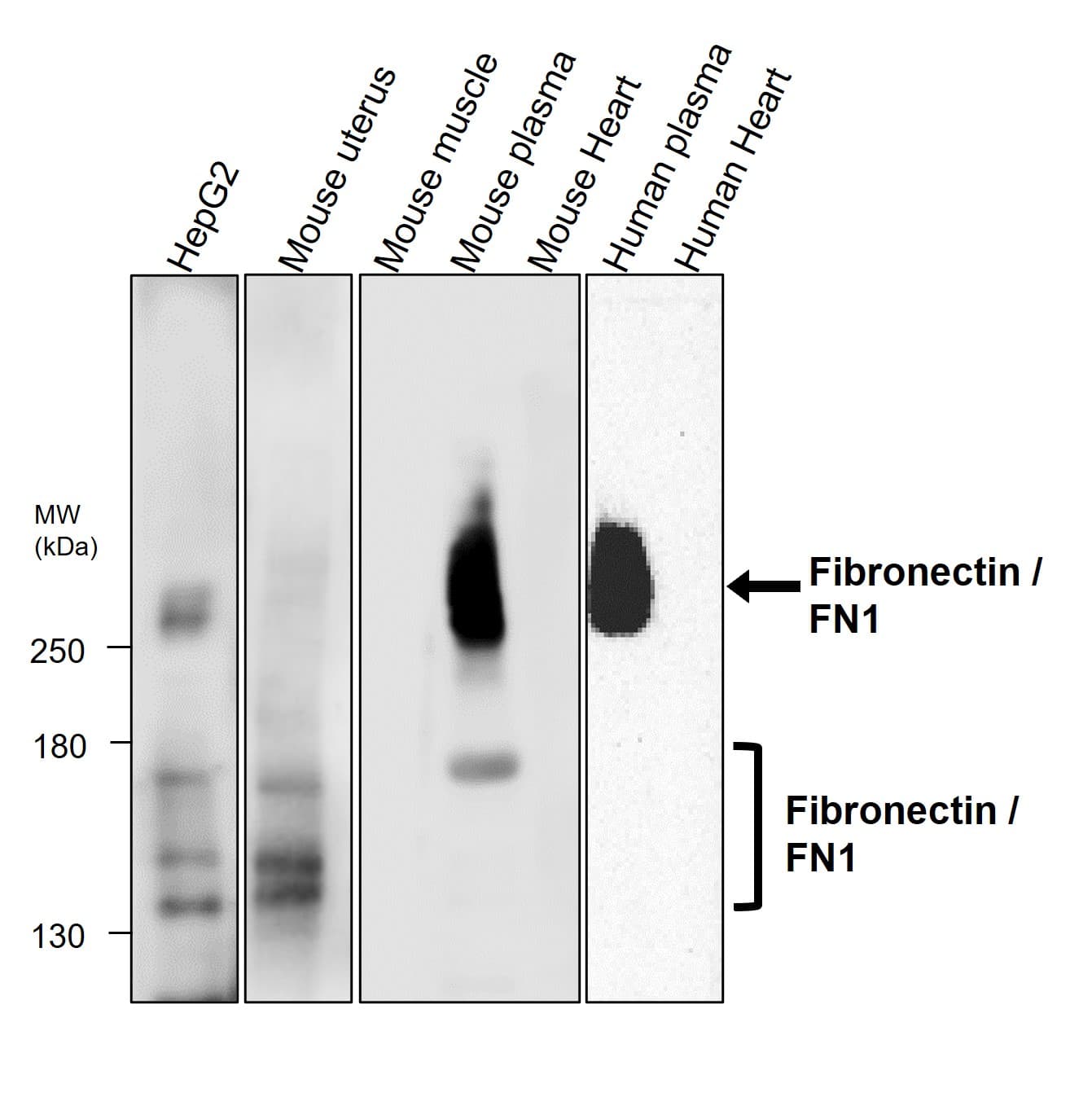
All lanes : Anti-Fibronectin antibody at 1/500 dilution. Lysates at 60 μg per lane. This blot was produced using a 5% SDS-PAGE. Nitrocellulose membrane was then blocked with 3%BSA for an hour before being incubated with IR48-138 overnight at 4°C. |
Immunohistochemical analysis of paraffin embedded human hepatocellular carcinoma (HCC) labeling Fibronectin with IR48-138 at 1/100.
|
|
|
IR48-138 anti-Fibronectin antibody IHC image |
IR48-138 anti-Fibronectin antibody IHC image |
|
|
|
|
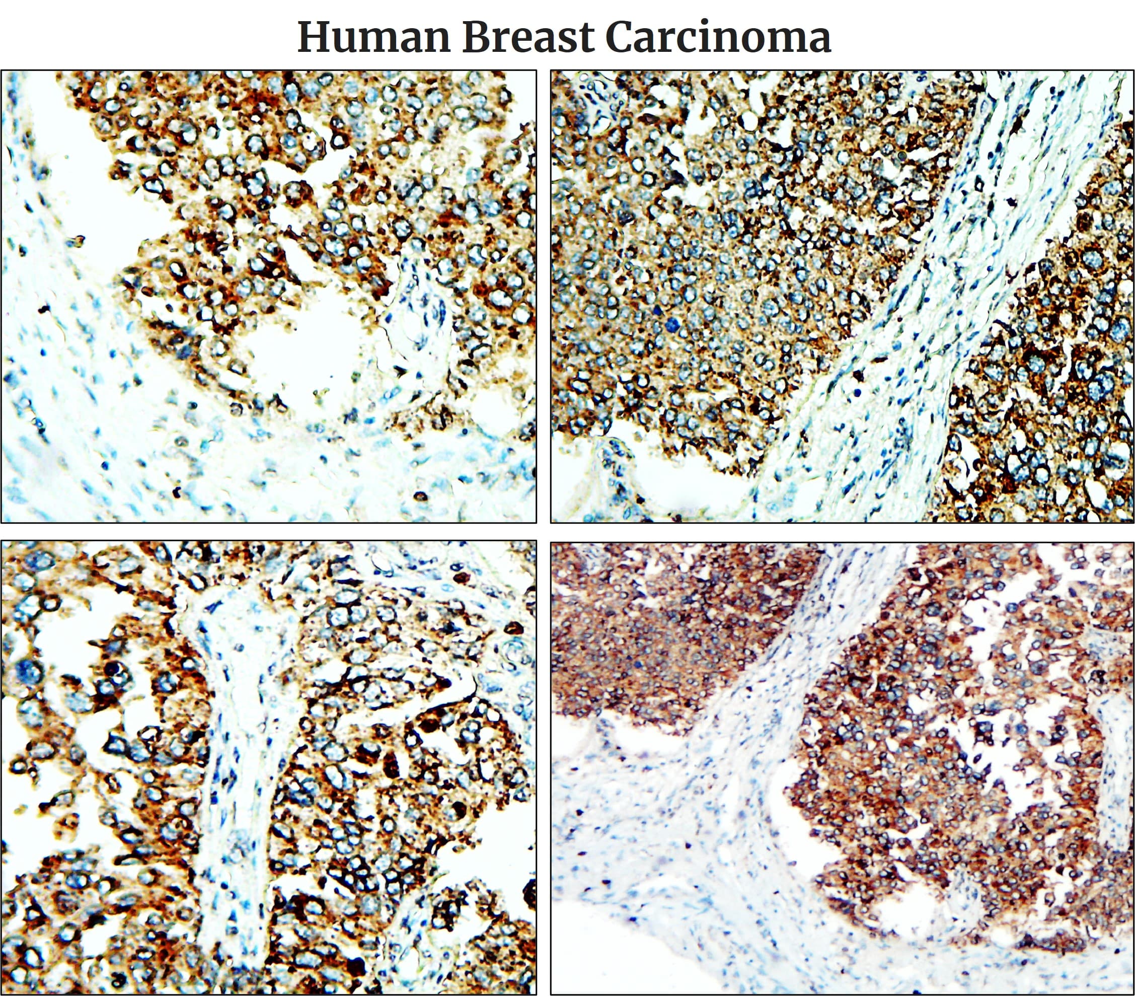
Immunohistochemical analysis of paraffin embedded human breast carcinoma labeling Fibronectin with IR48-138 at 1/100. |
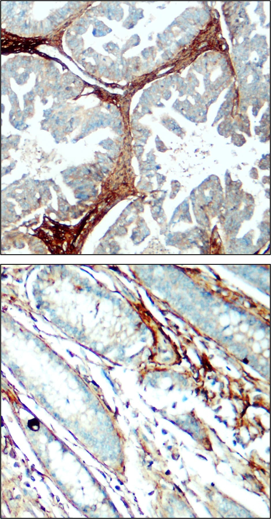
Immunohistochemical analysis of paraffin embedded Human cancer tissue labeling Fibronectin with IR48-138 at 1/100. |
|
Image |
|
|
|
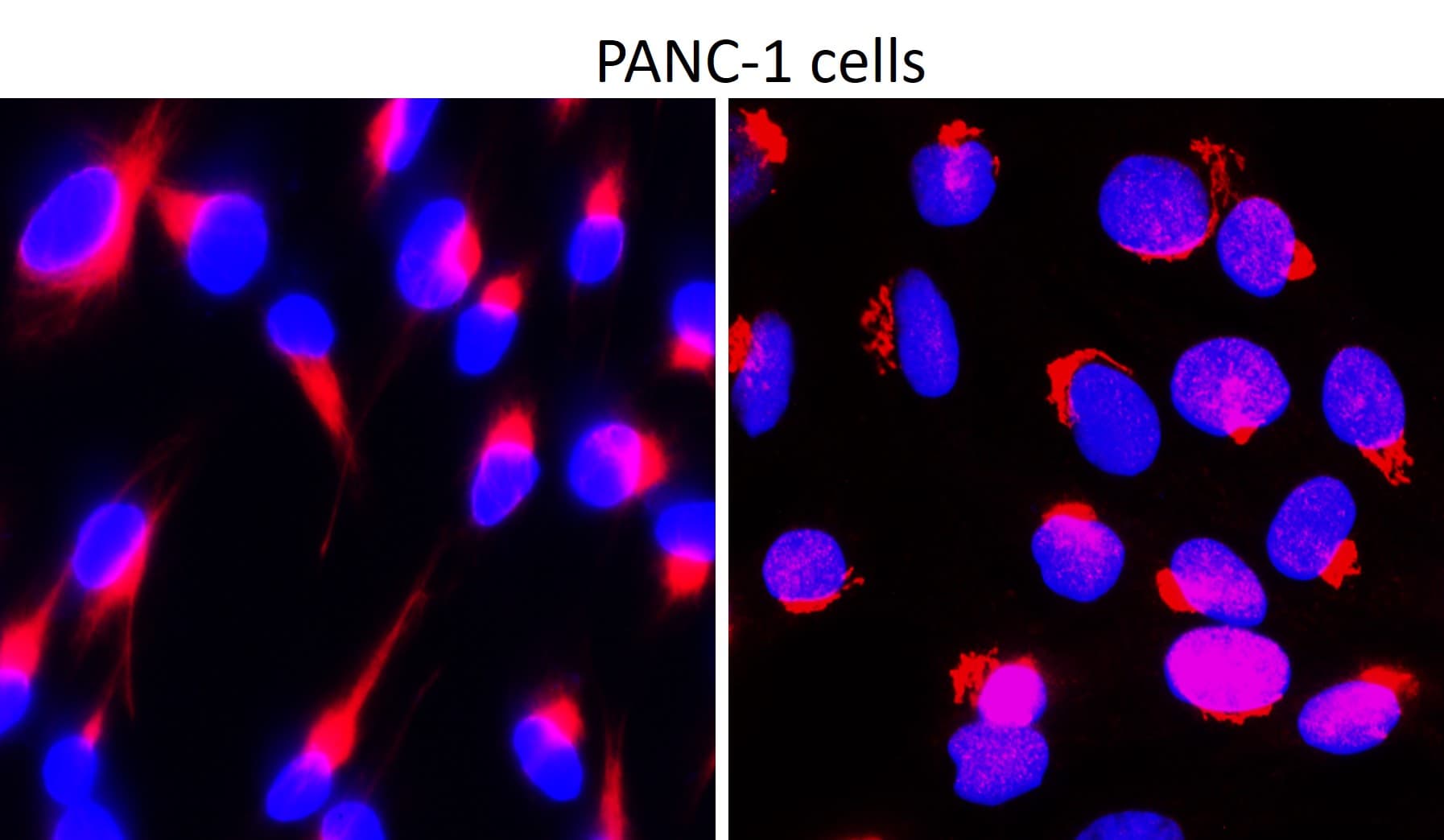
IR48-147 anti-Fibronectin antibody ICC/IF image |
|
Immunofluorescence: cells were fixed with 4% paraformaldehyde for 10 min at RT, permeabilized with 0.1% NP-40 for 10 min at RT then blocked with 5% BSA for 30 min at room temperature. Cells were stained with IR48-138 anti-Fibronectin antibody (red) at 1:200 and 4°C. DAPI (blue) was used as the nuclear counter stain. |
|